
একটি কোষের আত্মকাহিনী
Nusrat Surobhi
Catagories:
Academic articles,
PATHOLOGY
” জন্মিলে মরিতে হবে,
অমর কে কোথা কবে,
চিরস্থির কবে নীর, হায় রে, জীবন-নদে?” –
(বঙ্গভূমির প্রতি)
কবি মাইকেল মধুসূদন দত্তের লেখা এই লাইনগুলো আমাদের মনে করিয়ে দেয় যে, মানুষ মরণশীল। এই সুন্দর পৃথিবী ছেড়ে একদিন আমাদের সবারই পা বাড়াতে হবে অমোঘ যাত্রাপথে।
কি মনে হচ্ছে, আজকে সাহিত্যের দিকে চলে যাচ্ছি? 🤭
না না, আমরা প্যাথোলজিতেই থাকবো। 😁 উপরে মানুষের মরণশীলতা নিয়ে কথা বলার কারণ হলো, আজকে আমরা মানবদেহের একক cell এর মরণশীলতা নিয়ে কথা বলবো।
ঠিক ধরেছেন 😃। আমাদের আজকের টপিক হলো 👉 Cell Death.
💠আমাদের বডিতে প্রধানত 2 type এর cell death দেখা যায়।
1. Necrosis
2. Apoptosis
They differ in their mechanism, morphology, role in physiology & disease.
⭕ Necrosis : It is a pathological process that is the consequence of severe cell injury.
◾Necrosis কি কি কারণে হয় ❓
– Ischemia
– Exposure to microbial toxin
– Infection
– Proteolytic enzyme
– Trauma
🔶 Necrosis এ প্রধানত কি কি ঘটনা ঘটে ❓
1. Cellular protein গুলোর denaturation হয়।
2. Cellular content এর leakage হয়।
3. Locally inflammation দেখা দেয়।
4. যে cell গুলো lethally injured হয়ে গেছে সেগুলোর enzymatic digestion হয়।
✳️আচ্ছা, একটা cell এর severe injury হলো কিনা সেটা আমরা কিভাবে detect করতে পারি?
👉 Damage associated molecular pattern ( DAMPs) includes ATP, uric acid & other molecule which are normally confined within the healthy cell. But, their release indicate severe cell injury.
Damaged plasma membrane দিয়ে যদি intracellular protein গুলো leak করে circulation এ চলে আসে, তাহলে blood test এর মাধ্যমে জানা যাবে যে severe cell injury হয়েছে।
যেমন, Myocardial cell এর necrosis এর 2 hours এর মধ্যে blood এ Troponin – I detected হয়।
🔷 তো, এবারে আমরা জানবো, necrotic cell এর কি কি morphological change হয় ❓
🔺Increased eosinophilia :
Cytoplasmic RNA loss হবার কারণে cell এর acidity কমে যায়। আমরা জানি যে, always, acid attracts base. Acidity কমে গেলে, basophilia কমে যাবে। আর, denatured cytoplasmic protein accumulation হওয়ায় সেগুলো red stain eosin এর সাথে bind করে। Ultimately, eosinophilia বেড়ে যায়।
🔺 Glassy homogenous appearance due to loss of glycogen particles.
🔺Cytoplasm vaculolated হয়ে যায় এবং enzymatic digestion এর কারণে appearance দেখে মনে হয় যেন moth একটু একটু করে cytoplasm এর বিভিন্ন অংশ খেয়ে নিয়েছে 😶। তাই বলা হয়, moth-eaten appearance.
🔺Dead cell গুলো large, whorled phospholipid
mass এসে replace করে দেয় যাকে বলা হয় myelin figure.
🔺এই phospholipid গুলোর আবার degradation হয়ে fatty acid তৈরি হয় যা calcium এর সাথে bind করে calcium rich – precipitate deposit করে ফেলে।
💠 আমরা যেহেতু cell নিয়ে এতো পড়াশোনা করছি, তো necrosis হলে cell এর প্রাণকেন্দ্র nucleus এ কি কি change হয়, সেটাও তো অবশ্যই জানা দরকার তাই না?☺️
👉 🔴 Nuclear changes :- 3 টা pattern এ হয়।
1. Kariolysis
2.Pyknosis
3. Karryorrhexis
🔵 Kariyolysis:-
Endonuclease এর enzymatic degradation এর কারণে DNA loss হয় ➡ Chromatin fiber এর Basophilia কমে যায় ➡ Kariyolysis
🔵Pyknosis:-
Chromatin condenses into dense, shrunken basophilic mass ➡ Pyknosis.
🔵 Karryorrhexis:-
Pyknotic nucleus undergoes fragmentation ➡ nucleus of necrotic cell totally disappears ➡ It is called Karryorrhexis.
🔶 এবারে আমরা জেনে নেই necrosis এর কিছু pattern সম্পর্কে 💁♀️
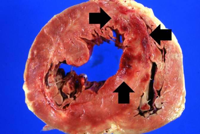
🔰 Coagulative necrosis:-
◾এই ক্ষেত্রে দেখা যায় যে, dead tissue এর architecture টা preserved হয় এবং firm texture থাকে।
◾Proteolysis is prevented by denaturation of structural protein & enzymes.
◾Necrotic cells are removed by phagocytosis & digestion of dead cell by lysosomal enzyme.
👉Example: Ischemia in obstructed blood vessel may lead to coagulative necrosis.
🔰Liquefactive necrosis:-
◾Digestion of dead cell, tissue becomes liquid viscous mass.
◾Necrotic material একসময় leukocyte এর উপস্থিতিতে creamy, yellow appearance দেখায় যাকে আমরা বলি Pus.
🔴Very Important ✅✅✅
CNS এর cell গুলোতে coagulating necrosis হয় না। Liquefactive necrosis হয়৷
🔰Gangrenous necrosis:-
◾এটা নরমালি lowre limb এ হয় যেখানে blood supply কমে গিয়েছে৷
Bacterial degradative enzyme attracts leucocyte & causes inflammation. এই অবস্থাকে বলা হয় wet gangrene.
🔰Caseous necrosis:–
◾এই নামটি Cheese শব্দ থেকে এসেছে। এক্ষেত্রে necrotic area তে firm, friable white appearance দেখা যায়।
◾Microscopicslly, necrotic area তে structureless collection of lysed cell & amorphous debris একটা distinctive inflammatory border এর ভেতরে enclosed থাকে। এই অবস্থাকে বলা হয় Granuloma.
◾Tuberculous infection এ এরকম পাওয়া যায়।
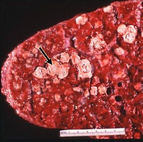
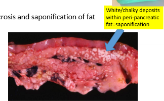
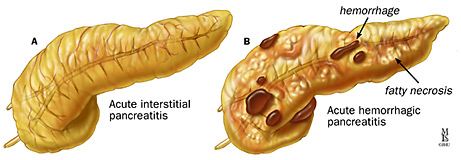
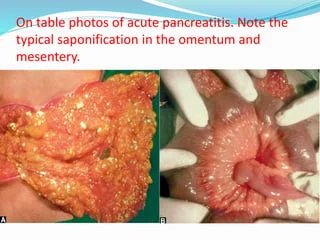
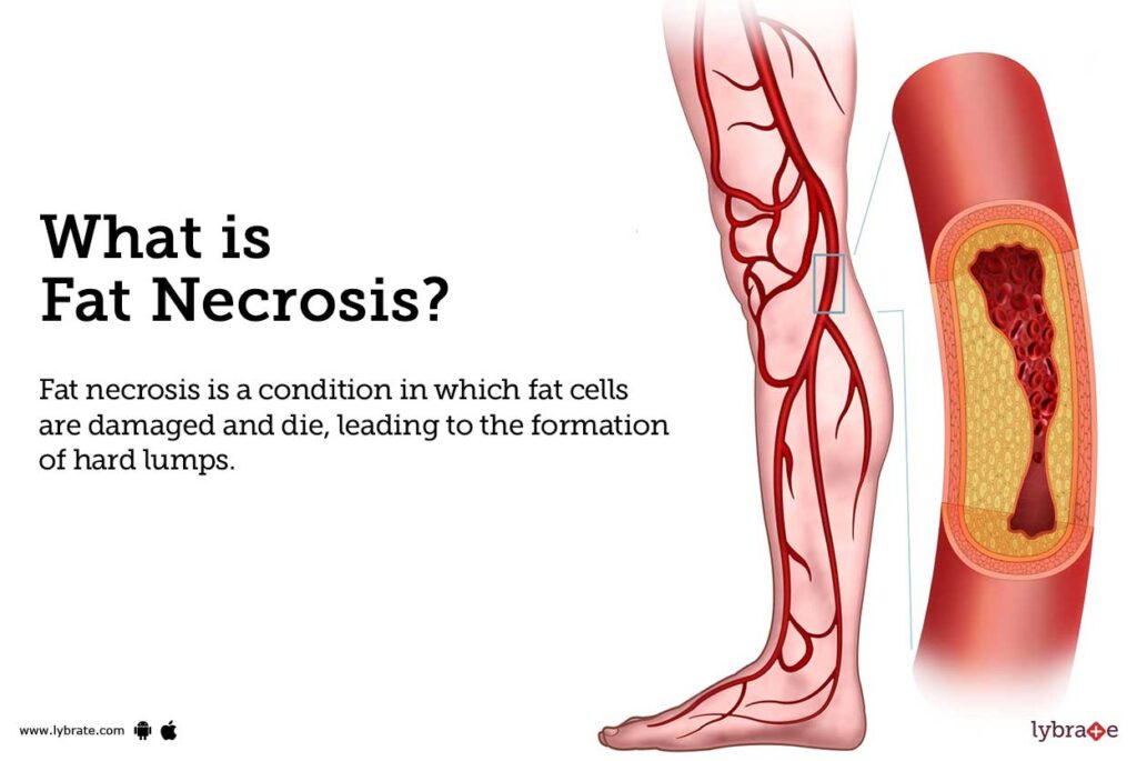
🔰Fat necrosis :-
আচ্ছা, আমরা কি কখনো ভেবে দেখেছি যে, pancreas এ যে এতো enzyme আছে, সেগুলো কখনো মুক্ত হয়ে peritoneal cavity তে মুক্ত হয়ে গেলে কি ঘটবে?🤔
◾Pancreatic lipase যখন peritoneal cavity & substance of pancreas এ চলে আসে, তখন fat cell এর membrane
liquefaction হয়ে যায়। যার ফলে triglyceride ester বের হয়ে আসে এবং pancreatic lipase তাকে ভেঙে Fatty acid produce করে।
◾Acute pancreatitis এ এরকম হয়।
◾এই Fatty acid তখন calcium এর সাথে bind করে grossly visible chalky white area তৈরি করে। একে বলে Fat saponification.
🔰Fibrinoid necrosis :-
◾কোনো vascular damage হলে arterial wall এ যদি antigen – antibody complex deposited হয় এবং এদের সাথে blood vessel থেকে leak করা plasma protein combined হয়ে যায়, তখন Hematoxylin & Eosin stain এ সেখানে bright pink appearance দেখা যায়। Pathologist রা এটাকে বলেন Fibrinoid necrosis.
💁♀️আমাদের আজকের পড়াশোনা necrosis পর্যন্তই থাকুক।
সবাই ভালো থাকুন, সুস্থ থাকুন । ধন্যবাদ সবাইকে। 😊
Nusrat Jahan Surobhi
Shaheed Suhrawardy Medical College
Session : 2018-19
Share this blog to social media:
Tags:
Suggested post
No post related to the current post. Please click on 'view more' to see more posts